


Author: Matthew Wells, DO
Description
A giant cell tumor of bone (GCT) is a bone tumor that is usually benign, which means it is not cancer. Even though it is not cancerous, it can still grow and damage the nearby bone if it is not treated. GCTs are most often seen in young adults between 20 and 40 years old and are slightly more common in women. These tumors usually develop near the ends of bones close to a joint, most commonly around the knee or wrist. Because they grow near joints, giant cell tumors can cause pain, swelling, stiffness, or trouble using the joint normally. If left untreated, the tumor can weaken the bone and, in some cases, increase the risk of a fracture.
Symptoms
The most common symptoms of a giant cell tumor are pain and swelling near a joint. Many patients describe the pain as a deep, ongoing ache that may get worse with activity. The area may feel tender to touch and may look swollen. Because these tumors often grow close to a joint, they can sometimes cause stiffness or make it harder to move the joint normally. In some cases, the tumor can weaken the bone enough to cause a fracture (broken bone), which may lead to sudden, severe pain.
Examination
During the physical exam, your doctor may notice tenderness or swelling near the affected bone, often close to a joint. The nearby joint may feel stiff or may not move as well as normal.
In some cases, your doctor may be able to feel a firm area or lump. Unlike an infection, the skin over the area is usually not red or warm.
Tests
X-rays are usually the first test used to diagnose a giant cell tumor of bone. On an X-ray, the tumor often appears as an area where the bone looks thinner or damaged, usually near the end of a bone close to a joint. Some tumors can be more aggressive and may cause the bone to expand or become very weak. A CT scan may be ordered to get a more detailed look at the bone and to help plan surgery. In some cases, an MRI is also needed to see if the tumor is close to important structures such as nerves or blood vessels. To confirm the diagnosis, a biopsy is needed. A biopsy involves taking a small sample of the tumor so it can be examined under a microscope. In many cases, the biopsy can be done during the same surgery used to treat the tumor.
Images
X-rays often show a giant cell tumor as an area where the bone has been worn away or weakened, usually near the end of a bone close to a joint. These tumors can sometimes cause the bone to expand or thin out as they grow (Figure 1).
In more advanced cases, the tumor may grow more aggressively and cause significant damage to the bone (Figure 2).
A CT scan is often used to get a more detailed view of the tumor and to better understand how much of the bone is involved. This information helps your surgeon plan the safest and most effective treatment (Figure 3).
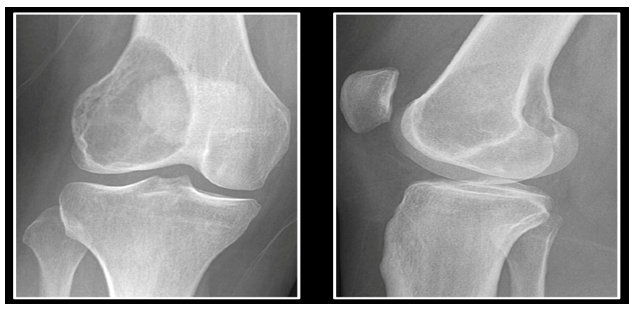
Figure 1. Anterior-posterior (AP) and lateral X-ray of a giant cell tumor within the distal femur demonstrating the typical lytic appearance near the joint surface.
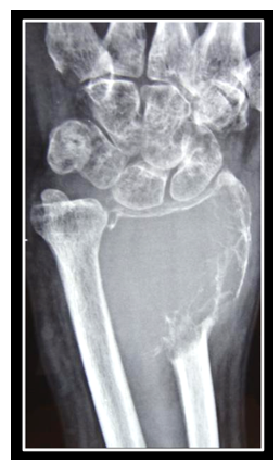
Figure 2. Anterior-posterior (AP) X-rays of a giant cell tumor within the distal radius demonstrating aggressive expansion with near complete erosion of the involved bone.
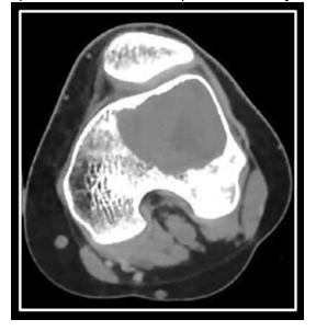
Figure 3. A computed tomography (CT) scan of the same distal femur from Figure 1 showing the giant cell tumor of bone extending to the subchondral (near-joint) bone with associated cortical thinning.
Prognosis
Giant cell tumors of bone are usually not life-threatening, but they can continue to grow and sometimes come back after treatment. About 1 in 4 to 5 patients may have the tumor return in the same area after surgery.
Surgery is the most common treatment. During surgery, the tumor is carefully removed by scraping it out of the bone (this is called curettage). The empty space left behind is then usually filled with bone graft or bone cement to help support the bone. In some cases, the bone may still be weak after surgery, so your surgeon may recommend adding metal plates, screws, or other support to help prevent the bone from breaking while it heals.
Your surgeon may also use additional treatments during surgery to lower the chance of the tumor coming back. For example, liquid nitrogen (freezing treatment) may be used to destroy any remaining tumor cells. In some patients, a medication may also be used that can help slow the growth of the tumor, strengthen the bone, and sometimes make surgery safer or easier, especially for tumors in difficult locations or when surgery needs to be delayed.
Because giant cell tumors of bone are uncommon and can sometimes behave aggressively or look similar to other bone tumors, evaluation by an orthopaedic oncologist is often recommended. Orthopaedic oncologists have specialized training in diagnosing and treating bone tumors, helping ensure an accurate diagnosis, careful surgical planning, and the best possible long-term outcome.
More Information:
- https://orthoinfo.aaos.org/en/diseases--conditions/giant-cell-tumor-of-bone
- https://www.cancer.gov/pediatric-adult-rare-tumor/rare-tumors/rare-bone-tumors/giant-cell-tumor-of-bone
- https://www.bcrt.org.uk/information/information-by-type/giant-cell-tumor/
- https://www.giantcelltumor.org/
This is not intended as a substitute for professional medical advice and does not provide advice on treatments or conditions for individual patients. All health and treatment decisions must be made in consultation with your physician(s), utilizing your specific medical information. Inclusion in this is not a recommendation of any product, treatment, physician or hospital.